 |
Did you know that micro- organisms come in all sorts of colors? From a warm red from a yeast like Sporobolomyces salmonicolor, fresh green of the algae Chlorella vulgaris, oily brown of the diatom Phaeodactylum tricornutum to even a vibrant violet as seen from the bacterium Rhodospirillum rubrum, microorganisms display a rainbow of colors. These colors are caused by special cell processes or by the creation of specific pigments. |
|||
| Bacteria | Cyanobacteria | Diatoms | Plasmids | |
Bacteria
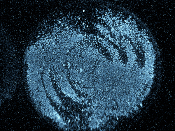
Aliivibrio fischeri (LMG 26186)
Aliivibrio fischeri is a bacterium ubiquitously distributed in sub-tropical and temperate marine environments. These bacteria emit blue light through a chemical reaction known as bioluminescence, which involves the oxidation of a molecule called luciferin in the presence of the enzyme luciferase.
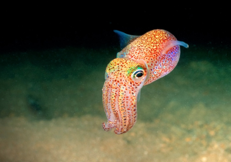
This bioluminescent species is commonly found in the light organs of various marine organisms, such as the Hawaiian bobtail squid. The light produced by this micro hero serves a variety of functions for the host organism, such as camouflage, attracting prey, and communication. by allowing the squid to match its body luminosity with the surrounding water, he avoids being detected by predators.
Cyanobacteria
Cyanobacteria are notable for their vibrant and diverse range of pigments, which have essential functions, contributing to their survival.
Chlorophyll
Chlorophyll is the most well-known pigment found in this group. Like in plants, it helps capturing light energy, converting it into chemical energy through photosynthesis, such as in this Nostoc (ULC 760) culture.
Carotenoids
Cyanobacteria also contain carotenoids, responsible for the orange/red/yellow hues often seen in blooms. These pigments not only aid in light absorption but also provide protection against excessive light. It can be observed in this culture of Calothrix (ULC 003).
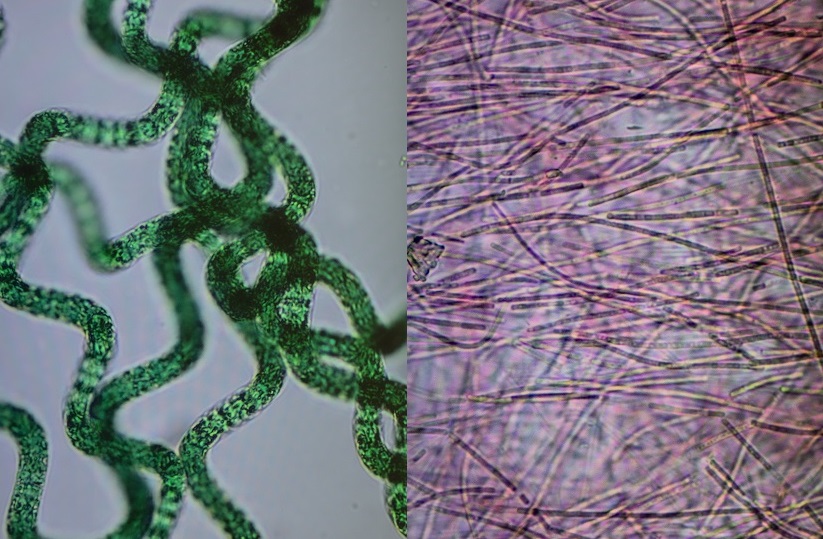
Phycobilins
Phycobilins are another set of pigments found in cyanobacteria. These include phycocyanin (blue in e.g Arthrospira, ULC 445, left), allophycocyanin (yellow/orange) and phycoerythrin (red in e.g. Leptolyngbya, ULC 764, right), which play a crucial role in capturing light energy and transferring it to chlorophyll molecules for photosynthesis.
The diversity of pigments in cyanobacteria serves not only as a visual spectacle but also as an adaptation that enables these microorganisms to inhabit a wide range of ecological niches. From freshwater to marine environments, and from surface waters to deep-sea thermal vents, cyanobacteria's pigments play a vital role in their ability to harness light energy, survive, and contribute to the planet's ecosystems.
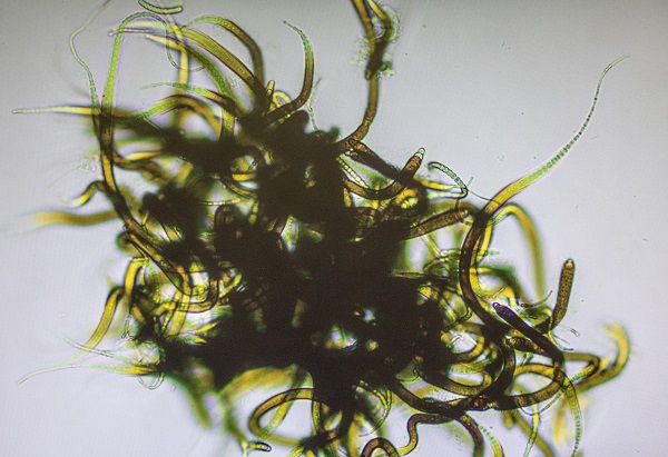
Diatoms and other microalgae
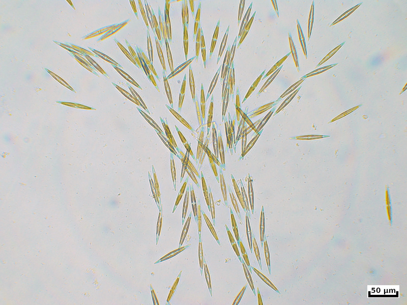
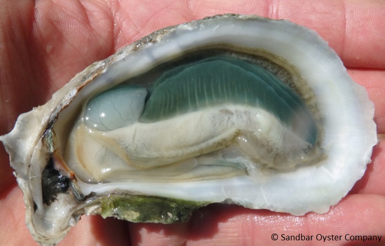
Haslea ostrearia (DCG 1054)
Green gilled oysters are regarded as a rare delicacy with a vibrant emerald colour and a special taste. This colouration is caused by a water-soluble blue pigment named marennine which is produced by the diatom Haslea ostrearia .
Haslea ostrearia is a thychopelagic/benthic diatom with a pennate shape. At least 4 other Haslea species have been discovered that form the marennine pigment (H. ostrearia, H. silbo, H. provincialis, H. nusantara) or a marennine-like pigment (H. karadagensis). They have been primarily found in the northern hemisphere. This unique blue pigment is of interest for many different branches varying from cosmetic purposes to cancer research.
A blue pigment that can be harvested from algae would seem more profitable than acquiring it from plants and having to deal with the remaining organic waste matter. However, large scale and sustainable cultivation of these diatoms can still be challenging. Safe for being a pretty colour, marennine also has proven to function as an antioxidant and could even function as a pH-indicator (blue = acidic, green = alkalic). Marennine can also react when coming into contact with biogenic amines originating from decomposing fish, such as putrescine, cadaverine, tyramine, dimethylamine and trimethylamine, rendering the pigment interesting as for food safety control.
The pigment can also act as an anti-proliferating agent and thus counteract cell growth. This has been successfully proven to work on a few cancer cell lines (e.g. melanoma, ovaries, colon, kidney, lung, breast). It has also proven to have antiviral properties against the HSV1 herpes virus and antibacterial properties against the Vibrio aestuarianus pathogen, but not a broad bacterial spectrum for human interests. However, it still plays a crucial role in the shaping of the microbiome surrounding Haslea ostrearia, as benthic diatoms and bacteria have a very close symbiotic relationship. This can also explain the absence of a variety of other microorganisms during Haslea blooms.
Harmful or harmless: Biological effects of marennine on marine organisms.
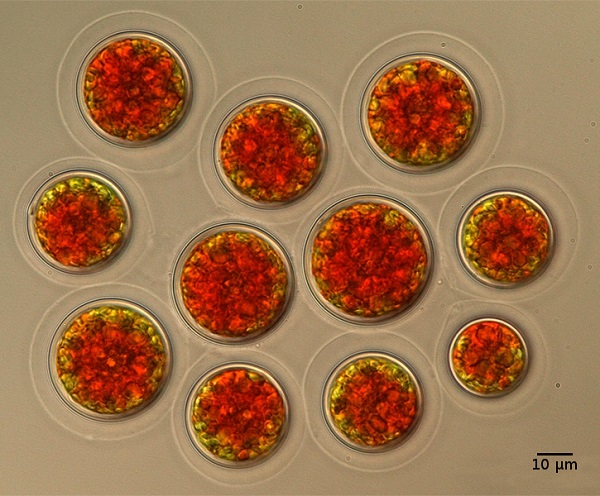
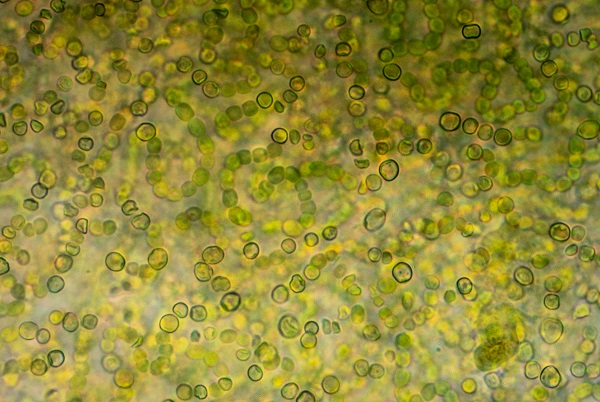
Haematococcus lacustris (DCG 0565)
Astaxanthin is a valuable red pigment produced by Haematococcus species. When in stressful environments, Haematococcus sp. forms resting spores which contain a lot of astaxanthin. When Haematococcus is consumed by shrimp, astaxanthin is incorporated into their exoskeleton, giving them a red color. The same applies to filterfeeding fish, as the astaxanthin collects in the meat of the fish, giving it a nice red color.
Did you know? Flamingos become pink due to their diet consisting of shrimp whose diet consists of Haematococcus algae!
Plasmids
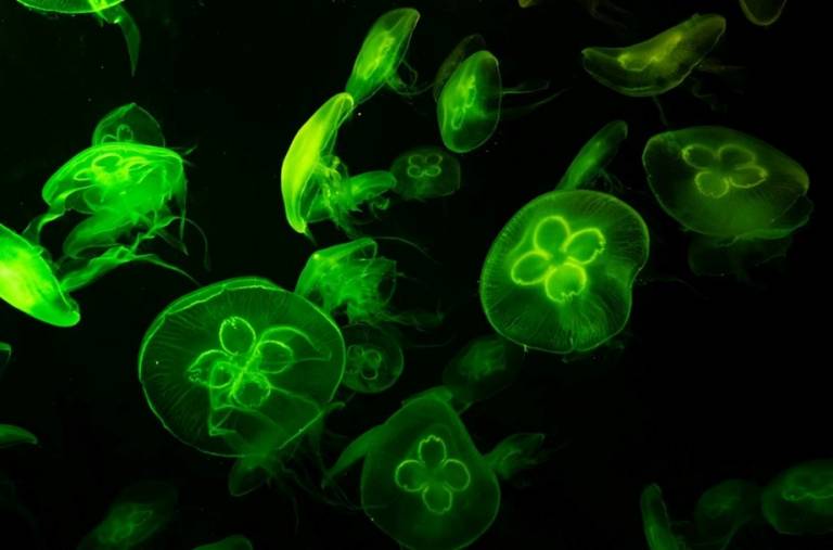
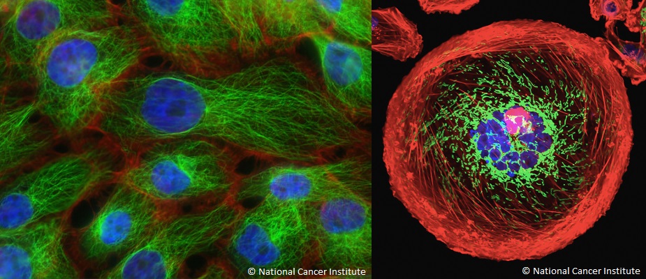
Green fluorescent protein (GFP)
Green fluorescent protein is a commonly used marker gene in biotechnology. It was originally isolated from the jellyfish Aequorea Victoria. When exposed to blue light, GFP will emit a bright green fluorescence.
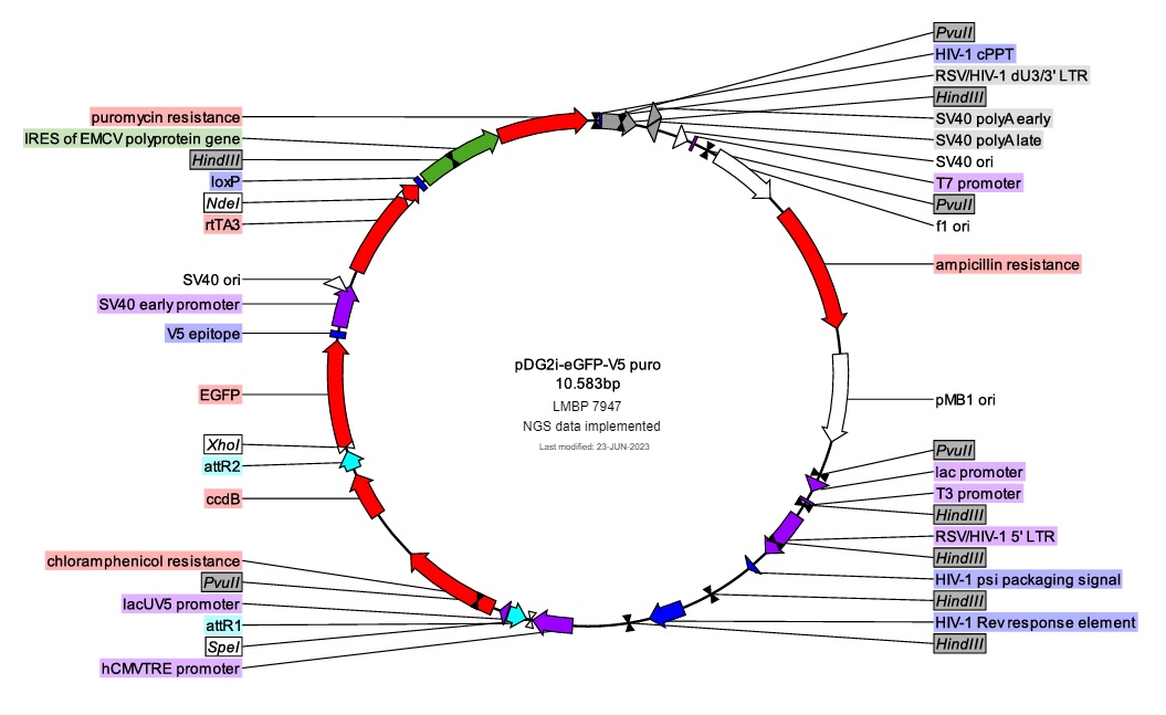
Scientists have created analogous proteins based on GFP, and later found and created other proteins with similar functions. Each of them emits a different colour after exposure to light of a specific wavelength. Through the use of plasmids, biotechnologists can create cells that express the proteins under study, fused to a fluorescent marker. Using several markers at the same time allows scientists to label different cell proteins simultaneously, each with a different fluorescent protein. The result is a colourful spectacle that shows us exactly how a cell is made and how cell components move over time.
In more complex experiments, various cell components can be marked via fluorescently labeled antibodies, which also means labeling is no longer limited to proteins. In this case, the result becomes even more breathtaking.